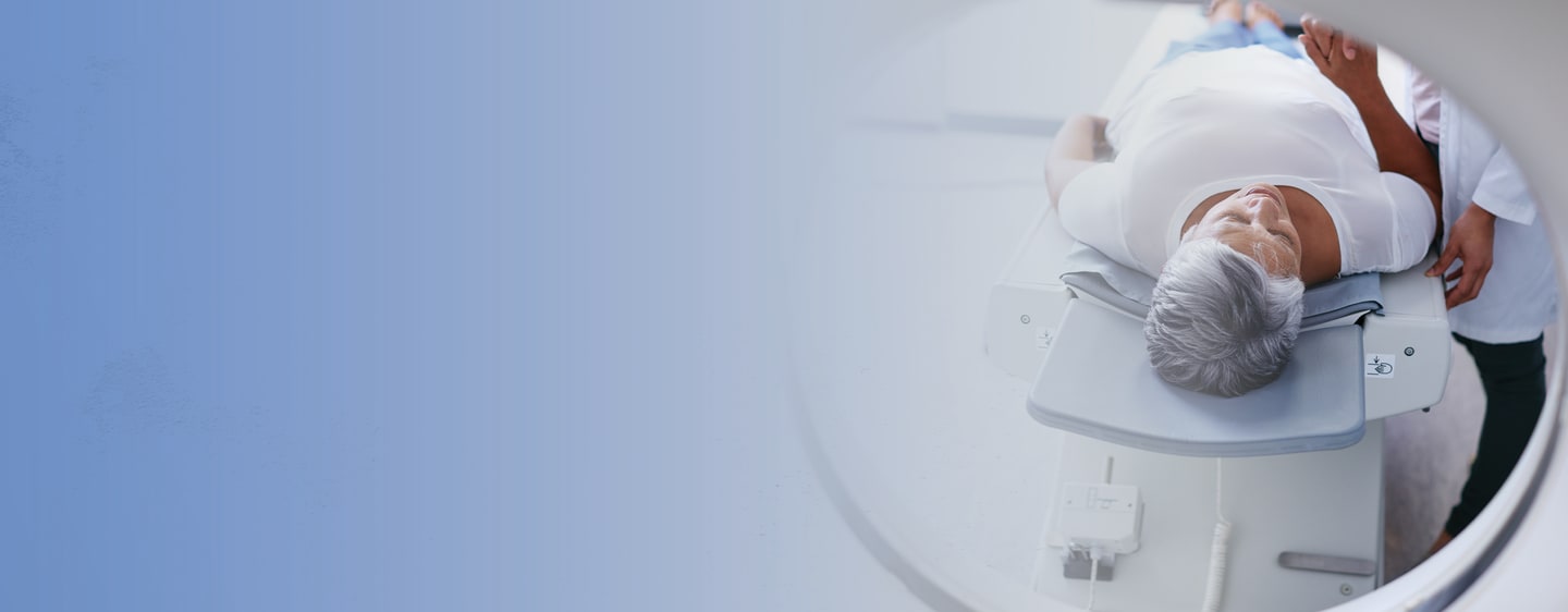
Diagnostic Imaging

Or, if you're not sure what you're looking for, you can:
Browse Specialists
Browse Primary Care
Or, if you're not sure what you're looking for, you can:
Browse All Conditions & Care Services


X-ray or radiography uses a very small dose of ionizing radiation to produce pictures of the body's internal structures. X-rays are the oldest and most frequently used form of medical imaging. They are often used to help diagnose fractured bones, look for injury or infection, and locate foreign soft-tissue objects. In addition, some x-ray exams may use an iodine-based contrast material or barium to help improve the visibility of specific organs, blood vessels, tissues, or bones.
A PET-CT scan combines a CT scan and a PET scan. This image can help pathologists find cancer and learn its stage. The CT scan combines a series of X-rays from all around your body to create a three-dimensional (3D) picture. The PET scan uses a mildly radioactive drug to highlight areas of your body where cells are more active than usual.
At Middlesex Health, our Radiology Department uses a cutting edge artificial intelligence program to improve the effectiveness of our PET-CT scans.
First, the AI program helps us reduce a patient's time lying on the scan table from 24 to 6 minutes. This allows for a more comfortable patient imaging experience while maintaining high-quality scans to help guide treatment.
In addition, the AI software alerts the radiologist to CT scans with critical imaging findings. As soon as the CT scan images are obtained, the software analyzes the images to detect urgent findings faster and reduce the time to start treatment.
Computed Tomography (CT) is a fast, painless, non-invasive, and accurate diagnostic imaging test that creates detailed images of internal organs, bones, soft tissue, and blood vessels. The cross-sectional images generated during a CT scan can be reformatted in multiple planes. They can even generate three-dimensional images. CT scanning is often the best method for detecting many different cancers since the images allow your doctor to confirm the presence of a tumor and determine its size and location. In emergency cases, it can reveal internal injuries and bleeding quickly enough to help save lives.
At Middlesex Health, our Radiology Department uses a cutting-edge artificial intelligence program to improve the effectiveness of our CT- scans.
This AI software alerts the radiologist to CT scans with critical imaging findings. As soon as the CT scan images are obtained, the software analyzes the images to detect brain bleeds, blood clots in the lungs, cervical spine fractures, and specific signs of a stroke. For patients being seen in the emergency department, this AI prioritization tool enables us to detect urgent findings faster and reduce the time to start treatment.
Fluoroscopy is a study of moving body structures - similar to an X-ray "movie." First, a continuous X-ray beam is passed through the body part being examined. Then, the beam is transmitted to a TV-like monitor to see the body part and its motion in detail.
Fluoroscopy is used in many types of imaging procedures. The most common uses of fluoroscopy include:
Barium swallow or barium enema: In these procedures, fluoroscopy is used to show the movement of the gastrointestinal (digestive) tract.
Cardiac catheterization: In this procedure, fluoroscopy shows blood flowing through the arteries. It is used to diagnose and treat some heart conditions.
Placement of catheter or stent inside the body: Catheters are thin, hollow tubes. They are used to get fluids into the body or to drain excess fluids from the body. Stents are devices that help open narrow or blocked blood vessels. Fluoroscopy helps ensure the proper placement of these devices.
Guidance in orthopedic surgery: A surgeon may use fluoroscopy to help guide procedures such as joint replacement and fracture (broken bone) repair.
Hysterosalpingogram: In this procedure, fluoroscopy is used to provide images of a woman's reproductive organs.
With Interventional Radiology (I/R), your doctor looks inside your body with imaging tests such as ultrasounds, CT scans, or MRIs. Then they use small tools, like needles and tubes, to do a procedure or give treatment right where you need it. The concept behind interventional radiology is to diagnose and treat patients using the least invasive techniques currently available to minimize risk to the patient and improve health outcomes. These procedures have less risk, less pain, and less recovery time in comparison to open surgery.
Nuclear Medicine uses very small amounts of radioactive material, either injected into a vein in the arm, inhaled, or swallowed, to diagnose and treat disease. Then, using a computer and a gamma camera to detect emitted radiation, images are produced that can determine cellular and organ function and structure.
Magnetic Resonance Imaging (MRI) uses a powerful magnetic field, radio waves, and a computer to produce detailed pictures of the body's internal structures that are clearer, more detailed, and more likely in some instances to identify and accurately characterize disease than other imaging methods. MRI's are used to evaluate the body for a variety of conditions, including tumors and diseases of the breast, liver, heart, and bowel. In addition, MRI's are non-invasive and do not use ionizing radiation.
Ultrasound imaging uses a transducer or probe to generate sound waves and produce pictures of the body's internal structures. It does not use ionizing radiation, has no known harmful effects, and provides a clear picture of soft tissues that don't appear well on x-ray images. Ultrasound is often used to help diagnose unexplained pain, swelling, and infection. It may also provide imaging guidance to needle biopsies or to see and evaluate conditions related to blood flow. It's also the preferred imaging method for monitoring a pregnant woman and her unborn child.
Bone Densitometry, also called dual-energy x-ray absorptiometry, DEXA, or DXA, is simple, quick, and non-invasive. The procedure uses a small dose of ionizing radiation to measure bone loss inside the body (usually the lower spine and hips). It is the most commonly used method for diagnosing osteoporosis and assessing an individual's risk for developing osteoporotic fractures.
In recognition of our commitment to providing the highest quality care, the Middlesex Health Diagnostic Imaging facilities in Middletown and Westbrook are designated as American College of Radiology (ACR) Comprehensive Breast Imaging Centers. Our Imaging facilites offer:
Lung cancer screening is a non-invasive test to look for nodules, or growths, in the lungs. It is performed using a low-dose CT scan and takes less than 30 minutes. Early-stage lung cancer usually does not have symptoms. However, for those with a high risk of lung cancer, screening can help your doctor detect the disease at stage 1 or stage 2 when it can be more easily treated.
Middlesex Health is proud to be a Lung Cancer Screening Center of Excellence. This designation is from the GO2 Foundation for Lung Cancer. It affirms our commitment to responsible, high-quality screening practices and compliance with professional medical organizations' best-practice standards.
A heart scan, also known as a Coronary Calcium Scan, is a specialized X-ray test that provides pictures of your heart to help your doctor detect and measure calcium-containing plaque in your arteries.
Plaque inside the arteries of your heart can grow and restrict blood flow to the muscles of your heart. Measuring calcified plaque with a heart scan may allow your doctor to identify possible coronary artery disease before you have signs and symptoms.
Your doctor will use your test results to determine what you need — medication or lifestyle changes — to reduce your risk of a heart attack or other heart problems.
Middlesex Health has partnered with the Mayo Clinic to bring advanced artificial intelligence technology to our diagnostic imaging offices. This new technology helps providers identify potential areas of concern, allowing for a more accurate diagnosis and getting you on the road to treatment and recovery faster.
Clark Otley, MD, Chief Medical Officer of The Mayo Clinic Platform, and Ravi Jain, MD, Ph.D., a Radiologist at Radiologic Associates of Middletown, discuss how Middlesex Health uses artificial intelligence to improve our diagnostic imaging practice.
Requesting a copy of your images is quick and simple. Just call 860-358-6506.