Discogram
Overview
A discogram, also called discography, is an imaging test used to look for the cause of back pain. A discogram might help your healthcare professional determine if a specific disk in your spine is causing your back pain.
Spinal disks are spongelike cushions between the bones of the spine, called vertebrae. During a discogram, dye is injected into the soft center of one or more disks. The injection sometimes reproduces the back pain.
The dye also moves into any cracks in the disk's exterior, which can then be seen on an X-ray or CT scan. However, disks that show wear and tear don't always cause symptoms, so the usefulness of a discogram is debatable.
Why it's done
A discogram is an invasive test that generally isn't used for an initial exam of back pain. Your healthcare professional might suggest a discogram if your back pain persists despite conservative treatments, such as medicine and physical therapy.
Some healthcare professionals use a discogram before spinal fusion surgery to help identify which disks need to be removed. However, discograms are not always correct in identifying which disks, if any, are causing back pain. Many healthcare professionals instead rely on other tests, such as MRI and CT scanning, to diagnose disk problems and guide treatment.
Risks
A discogram is generally safe. But as with any medical procedure, a discogram carries a risk of complications, including:
- Infection.
- Worsening of chronic back pain.
- Headache.
- Injury to nerves or blood vessels in and around the spine.
- Allergic reaction to the dye.
How you prepare
You might need to stop taking blood-thinning medicines for a time before the procedure. Your healthcare team will tell you what medicines you can take. You will not eat or drink the morning before the test.
What you can expect
A discogram is performed in a clinic or hospital room that has imaging equipment. You'll likely be there for up to three hours. The test itself takes 30 to 60 minutes, depending on how many disks are examined.
Before the procedure
Although you're awake during the procedure, you might receive a medicine to help you feel calm or less anxious. You also might be given an antibiotic to help prevent infection.
During the procedure
You will lie on a table on your stomach or side. After cleaning your skin, a member of your healthcare team may inject a numbing medicine to decrease pain caused by the insertion of the discogram needle.
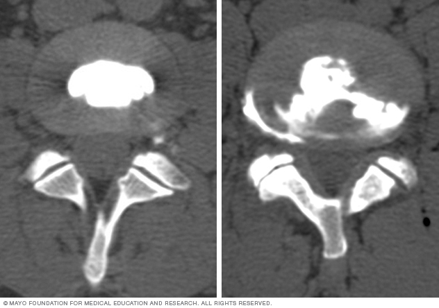
Your healthcare team will use an imaging technique called fluoroscopy to watch the discogram needle enter your body. Fluoroscopy allows more-precise and safer placement of the needle into the center of the disk to be examined. A contrast dye is then injected into the disk. Next, an X-ray or CT scan is taken to see if the dye spreads.
If the dye stays in the center of the disk, the disk is not damaged. If the dye spreads outside the center of the disk, the disk has undergone some wear-and-tear change. The change might or might not be the cause of your pain.
Typically, if a disk is causing your back pain, you will feel pain during the injection that's similar to the back pain you have daily. If a disk is not causing pain, there's little pain during the injection. During the discogram, you'll be asked to describe and rate your pain.
After the procedure
You will remain in the procedure room for approximately 30 to 60 minutes for observation. After that, you'll be able to go home. Someone will need to drive you.
Many people have some pain at the injection site or in the low back for several hours after the procedure. Applying an ice pack to the area for 20 minutes at a time might help. You'll need to keep your back dry for 24 hours.
If you develop severe back pain or a fever 1 to 2 weeks after the procedure, call your healthcare professional right away.
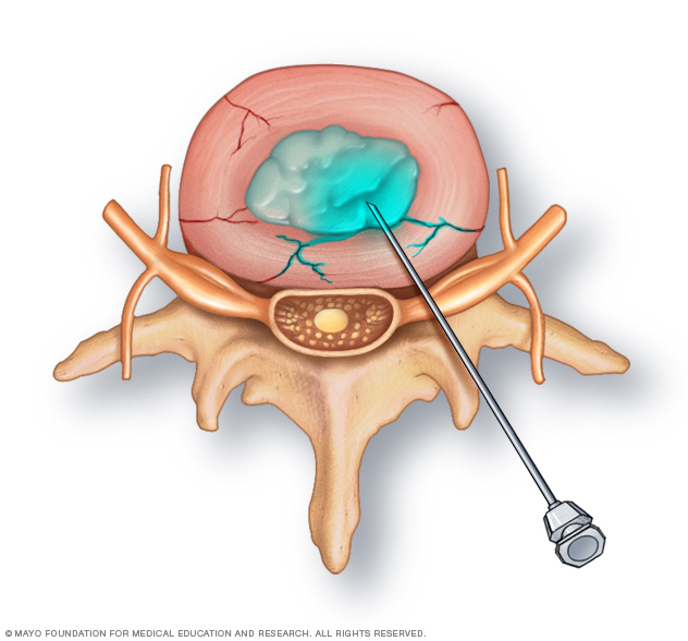
Your healthcare team inserts the tip of a needle into the center of each disk to be examined. A contrast dye is then injected into the disk.
If the dye stays in the center of the disk, the disk is not damaged. If the dye spreads outside the center of the disk, the disk has undergone wear-and-tear change, which may or may not be painful.
Results
Your healthcare professional will review the images and the information you provided about the pain you had during the procedure. This information will help your healthcare professional pinpoint the source of your back pain. Your healthcare team will use this information to guide your treatment or prepare for surgery.
Healthcare professionals usually don't rely on the results of a discogram alone because a disk with wear-and-tear change might not cause pain. Also, pain responses during a discogram can vary widely.
Often, results of a discogram are combined with results of other tests — such as an MRI or CT scan and physical exam — when determining a treatment plan for back pain.
Last Updated Apr 16, 2024
© 2024 Mayo Foundation for Medical Education and Research (MFMER). All rights reserved. Terms of Use