Breast lump or breast changes: Early evaluation is essential
Finding a breast lump or other change in a breast might cause worry about breast cancer.
That's understandable. But breast lumps are common. Most often they're noncancerous (benign), particularly in younger women. Still, it's important to have any breast lump evaluated by a health care provider, especially if it's new or if one breast feels different from the other breast.
How breast tissue feels
Breasts contain tissues of different textures, including fat, glands and connective tissue. Some breast-related symptoms, such as tenderness or lumpiness, change with the menstrual cycle. Lumps during this time might be caused by extra fluid in the breasts. Breast tissue also changes during pregnancy and menopause and while taking hormones.
When to consult a health care provider
Being familiar with how breasts usually feel makes it easier to detect when there's a change.
Reasons to consult a health care provider include:
- Finding a new breast lump or thickening that feels different from the surrounding tissue or the other breast
- Noticing a change in the size, shape or appearance of a breast
- Having breast pain that doesn't go away after the next period
- Noticing skin changes on a breast, such as itchiness, redness, scaling, dimpling or puckering
- Having a newly inverted nipple
- Noticing nipple discharge
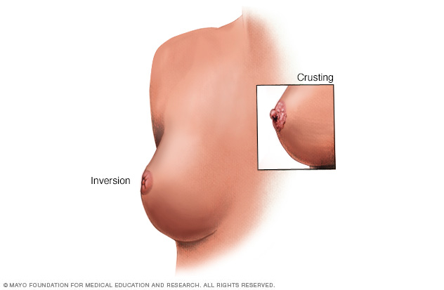
Breast and nipple changes can be a sign of breast cancer. Make an appointment with your health care provider if you notice anything unusual.
What to expect during a clinical breast exam
Evaluation of a breast lump typically begins with a clinical breast exam. During this exam, a health care provider will likely:
- Ask about symptoms and risk factors for breast cancer or benign breast conditions
- Examine the breasts and lymph nodes in the armpit, feeling for lumps or any other differences
- Examine the skin on the breasts
- Check for nipple problems, such as inversion or discharge
If the care provider finds a breast lump or other area of concern, you'll likely need testing.
Procedures to evaluate a breast lump
Imaging tests
To further evaluate a breast lump, a care provider might recommend:
- Diagnostic mammogram. This specialized breast X-ray shows breast changes. It takes X-ray pictures from several angles.
- Breast ultrasound. Sound waves create images of the inside of the breast on a monitor. Ultrasound imaging is helpful for determining whether a breast lump is solid or filled with fluid.
- Breast MRI. An MRI machine uses a magnet and radio waves to create pictures of the interior of a breast. A breast MRI usually is reserved for when the diagnosis is in question. Before a breast MRI, a dye might be injected through an intravenous (IV) line in an arm to enhance the appearance of tissues or blood vessels on the MRI pictures.
Newer tests for breast imaging are being developed and studied.
Breast biopsy
This involves having a tissue sample removed and examined under a microscope (biopsy). Ultrasound or mammography might help guide the needle, and a local anesthetic might be used. Breast biopsy options include:
- Fine-needle aspiration biopsy. With a thin needle attached to a syringe, cells and fluid are removed from the suspicious area.
- Core needle biopsy. A larger needle with a special tip is used to remove a sample of breast tissue.
- Stereotactic biopsy. Mammography produces images of the area in question from several angles (stereo images). A health care provider then removes a sample of breast tissue with a needle.
- Vacuum-assisted biopsy. A probe connected to a vacuum device removes a small sample of breast tissue.
- Surgical excisional biopsy. A small cut is made in the skin and breast tissue to remove part or all of a lump.
After a biopsy, the tissue sample is sent to a lab for analysis. A health care provider explains the results.
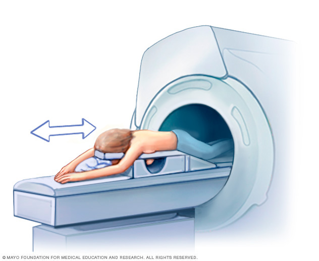
A breast MRI requires lying face down on a padded scanning table. The breasts fit into a hollow depression in the table, which contains coils that detect magnetic signals. The table slides into the large opening of the MRI machine.

During fine-needle aspiration, a special needle is inserted into a breast lump and any fluid is removed (aspirated). Ultrasound — a procedure that uses sound waves to create images of the breast on a monitor — might be used to help place the needle.

A core needle biopsy uses a long, hollow tube to take a sample of tissue. Here, a biopsy of a suspicious breast lump is being done. The sample is sent to a laboratory for testing and evaluation by doctors who specialize in analyzing blood and body tissue (pathologists).
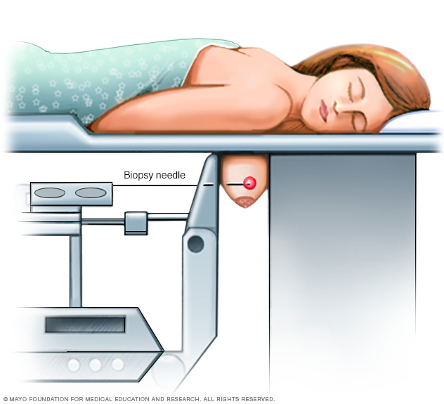
During a stereotactic breast biopsy, the breast is firmly compressed between two plates. X-rays (mammograms) are used to produce images of the same area from different angles. This is to determine the exact location for the biopsy. A sample of breast tissue in the area of concern is then removed with a needle.
Follow-up after breast lump evaluation
If the breast lump isn't cancerous, a health care provider will decide if there's a need for short-term monitoring with clinical breast exams or repeat breast imaging. You may be asked to return in 2 to 3 months to see if there have been changes in your breast. Consult your provider if you notice changes in the lump or develop new areas of concern.
A diagnosis in question might result in further consultation with a surgeon or other specialist. A diagnosis might be in question, for example, when a clinical breast exam and the mammogram show areas of suspicion, but the biopsy is benign.
A cancerous breast lump requires treatment. The tumor type and other factors will influence treatment options.
Last Updated Dec 3, 2022
© 2024 Mayo Foundation for Medical Education and Research (MFMER). All rights reserved. Terms of Use


