Aortic valve stenosis
Overview
Aortic valve stenosis — or aortic stenosis — is a type of heart valve disease (valvular heart disease). The valve between the lower left heart chamber and the body's main artery (aorta) is narrowed and doesn't open fully. This reduces or blocks blood flow from the heart to the aorta and to the rest of the body.
Treatment of aortic stenosis depends on the severity of the condition. You may need surgery to repair or replace the valve. Without treatment, severe aortic valve stenosis can lead to death.
Symptoms
Aortic valve stenosis ranges from mild to severe. Symptoms generally occur when narrowing of the valve is severe. Some people with aortic valve stenosis may not have symptoms for many years.
Symptoms of aortic valve stenosis may include:
- An irregular heart sound (heart murmur) heard through a stethoscope
- Chest pain (angina) or tightness with activity
- Feeling faint or dizzy or fainting with activity
- Shortness of breath, especially with activity
- Fatigue, especially during times of increased activity
- Rapid, fluttering heartbeat (palpitations)
- Not eating enough (mainly in children with aortic valve stenosis)
- Not gaining enough weight (mainly in children with aortic valve stenosis)
Aortic valve stenosis may lead to heart failure. Heart failure symptoms include fatigue, shortness of breath, and swollen ankles and feet.
When to see a doctor
If you develop symptoms that may suggest aortic valve stenosis, make an appointment with your health care provider.
Causes
To understand the causes of aortic valve stenosis, it may be helpful to know how the heart and heart valves typically work.
The heart has four valves that keep blood flowing in the correct direction:
- Aortic valve
- Mitral valve
- Tricuspid valve
- Pulmonary valve
Each valve has flaps (cusps or leaflets) that open and close once during each heartbeat. Sometimes, the valves don't open or close properly. If a valve doesn't fully open or close, blood flow is reduced or blocked.
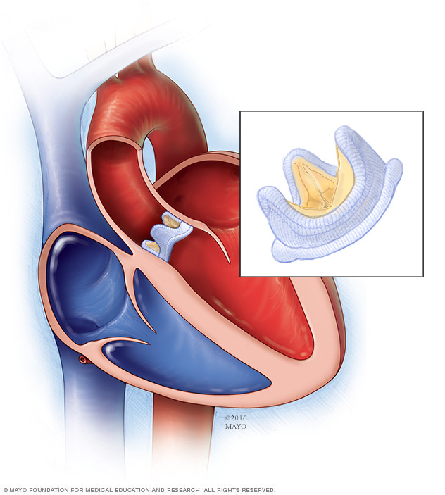
In aortic valve stenosis, the valve between the lower left heart chamber (left ventricle) and the aorta does not open completely. The area through which blood moves out of the heart to the aorta is narrowed (stenosis).
When the aortic valve opening is narrowed, the heart must work harder to pump enough blood into the aorta and to the rest of the body. The extra work of the heart can cause the left ventricle to thicken and enlarge. Eventually the strain can cause a weakened heart muscle and can ultimately lead to heart failure and other serious problems.
Aortic valve stenosis causes include:
-
Congenital heart defect. Some children are born with an aortic valve that has only two cusps (bicuspid aortic valve) instead of three (tricuspid aortic valve). Rarely, an aortic valve may have one (unicuspid) or four (quadricuspid) cusps.
Having a congenital heart defect such as a bicuspid aortic valve requires regular medical checkups. The valve condition may not cause any problems until adulthood. If the valve begins to narrow or leak, it may need to be repaired or replaced.
-
Calcium buildup on the valve (aortic valve calcification). Calcium is a mineral found in the blood. As blood repeatedly flows over the aortic valve, calcium deposits can build up on the heart valves.
The calcium deposits may never cause any problems. Aortic valve stenosis that's related to increasing age and calcium deposit buildup usually doesn't cause symptoms until age 70 or 80. However, in some people — particularly those with congenital aortic valve defects — calcium deposits result in stiffening of the valve cusps at a younger age.
-
Rheumatic fever. This complication of untreated strep throat can damage the heart valves. It may cause scar tissue to form on the aortic valve. Scar tissue can narrow the aortic valve opening or create a rough surface on which calcium deposits can collect.
Rheumatic fever may damage more than one heart valve, and in more than one way. While rheumatic fever is rare in the United States, some older adults had rheumatic fever as children.
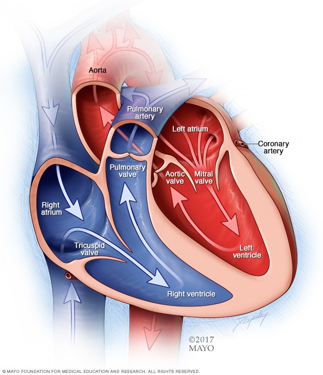
A typical heart has two upper and two lower chambers. The upper chambers, the right and left atria, receive incoming blood. The lower chambers, the more muscular right and left ventricles, pump blood out of the heart. The heart valves are gates at the chamber openings. They keep blood flowing in the right direction.

In aortic valve stenosis, the aortic valve opening is narrowed, as seen in the top image. The heart must work harder to pump blood across the smaller opening. This increases pressure within the heart. Eventually the strain reduces the heart's ability to pump blood to the body. This is like placing smaller and smaller nozzles on the end of a garden hose, as shown on the bottom image. The narrower the nozzle is, the slower the flow of water. This results in pressure buildup within the garden hose.
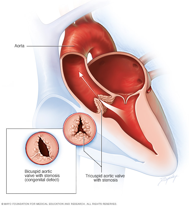
Aortic valve stenosis is a thickening and narrowing of the valve between the heart's main pumping chamber and the body's main artery, called the aorta. The narrowing creates a smaller opening for blood to pass through. This reduces or blocks blood flow from the heart to the rest of the body. Typically the aortic valve has three cusps, called a tricuspid aortic valve. But some people are born with an aortic valve that has two cusps, a condition called bicuspid aortic valve.
Risk factors
Risk factors of aortic valve stenosis include:
- Older age
- Certain heart conditions present at birth (congenital heart defects), such as a bicuspid aortic valve
- Chronic kidney disease
- Having heart disease risk factors, such as diabetes, high cholesterol and high blood pressure
- History of infections that can affect the heart, such as rheumatic fever and infective endocarditis
- History of radiation therapy to the chest
Complications
Aortic valve stenosis can cause complications, including:
- Heart failure
- Stroke
- Blood clots
- Bleeding
- Irregular heart rhythms (arrhythmias)
- Infections that affect the heart, such as endocarditis
- Death
Prevention
Some possible ways to prevent aortic valve stenosis include:
- Taking steps to prevent rheumatic fever. See your health care provider when you have a sore throat. Strep throat can usually be easily treated with antibiotics. Untreated strep throat can develop into rheumatic fever. Rheumatic fever is more common in children and young adults.
- Keeping the heart healthy. Talk to your health care provider about risk factors for heart disease and how to prevent or manage them. They include high blood pressure, obesity and high cholesterol levels. These risk factors may be linked to aortic valve stenosis.
- Taking care of the teeth and gums. There may be a link between infected gums (gingivitis) and infected heart tissue (endocarditis). Inflammation of heart tissue caused by infection can narrow arteries and worsen aortic valve stenosis.
If you have aortic valve stenosis, your health care provider may recommend that you limit strenuous activity to avoid overworking your heart.
Diagnosis
To diagnose aortic valve stenosis, your health care provider will examine you and ask questions about your symptoms and medical history. The provider will listen to your heart with a stethoscope to determine if you have a heart murmur related to an aortic valve condition.
Tests
Your health care provider may order several tests to confirm or rule out aortic valve stenosis. Tests also can help determine a cause and the condition's severity.
Tests for aortic valve stenosis may include:
-
Echocardiogram. An echocardiogram uses sound waves to create pictures of the beating heart. It shows how blood flows through the heart and heart valves. It can help identify a weakened heart muscle and determine the severity of aortic valve stenosis.
Sometimes, a special type of echocardiogram called transesophageal echocardiogram (TEE) may be done to get a closer look at the aortic valve. In this test, a flexible tube containing the ultrasound probe is guided down the throat and into the esophagus and placed closed to the heart.
- Electrocardiogram (ECG or EKG). This painless test measures the electrical activity of the heart. During an ECG, sensors (electrodes) are attached to the chest and sometimes to the arms or legs. An ECG can show how fast or slow the heart is beating. Your health care provider can look for signal patterns related to heart disease or swelling of the heart's chambers.
- Chest X-ray. A chest X-ray shows the condition of the heart and lungs. It can help determine whether the heart is enlarged, which can occur in aortic valve stenosis. It can also show swelling of the aorta and calcium buildup on the aortic valve.
- Exercise tests or stress tests. These tests often involve walking on a treadmill or riding a stationary bike while the heart is monitored. Exercise tests help determine whether symptoms of aortic valve disease occur during physical activity. If you're unable to exercise, medications that have similar effects as exercise on your heart may be used.
- Cardiac computerized tomography (CT) scan. A cardiac CT scan combines several X-ray images to provide a more detailed cross-sectional view of the heart. This test can help measure the size of the aorta and provide details about the aortic valve.
- Cardiac magnetic resonance imaging (MRI) scan. A cardiac MRI uses magnetic fields and radio waves to create detailed images of the heart. This test can show the size of the aorta. It may be used to determine the severity of aortic valve stenosis.
-
Cardiac catheterization. This test isn't often used to diagnose aortic valve disease, but it may be used if other tests aren't able to diagnose the condition or to determine its severity. It may also be used before aortic valve surgery to make sure the arteries that feed the heart muscle (coronary arteries) are not blocked.
In this procedure, a long, thin flexible tube (catheter) is inserted in a blood vessel, usually in the groin or wrist, and guided to the heart. Dye flows through the catheter to arteries in the heart. This is called a coronary angiogram. The dye helps the arteries show up more clearly on X-ray images and video. During the test, the pressure inside the heart chambers can be measured.
After testing confirms a diagnosis of aortic valve disease, your health care provider may tell you the stage of disease. Staging helps determine the most appropriate treatment.
Heart valve disease is staged into four basic groups:
- Stage A: At risk. Risk factors for heart valve disease are present.
- Stage B: Progressive. Valve disease is mild or moderate. There are no heart valve symptoms.
- Stage C: Asymptomatic severe. There are no heart valve symptoms but the valve disease is severe.
- Stage D: Symptomatic severe. Heart valve disease is severe and is causing symptoms.
The stage of heart valve disease depends on many things, including symptoms, disease severity, the structure of the valve or valves, and blood flow through the heart and lungs.
Treatment
Treatment for aortic valve stenosis depends on the symptoms and the severity of the condition.
If you have mild aortic valve symptoms or none, you may only need regular checkups by a health care provider. The provider may recommend healthy lifestyle changes and medications to treat valve disease symptoms or reduce the risk of complications.
Surgery or other procedures
You may eventually need surgery to repair or replace the diseased aortic valve, even if you don't have symptoms. Aortic valve surgery may be done at the same time as other heart surgery.
Surgery to repair or replace an aortic valve is usually done through a cut (incision) in the chest. Less invasive approaches may be available. Ask your health care provider which type of procedure is best for you. Aortic valve surgery may be done at the same time as other heart surgery.
Surgery options for aortic valve stenosis include:
-
Balloon valvuloplasty. This procedure can treat aortic valve stenosis in infants and children. In adults, the aortic valve tends to narrow again after the procedure. So it's usually only done if you're too ill for surgery or you're waiting for a valve replacement, because additional procedures are typically needed to treat the narrowed valve over time.
In this procedure, a long, thin tube (catheter) with a balloon on the tip is inserted into an artery in the arm or groin. It's guided to the aortic valve. Once in place, the balloon is inflated, which widens the valve opening. The balloon is then deflated, and the catheter and balloon are removed.
-
Aortic valve replacement. Aortic valve replacement is often needed to treat aortic valve stenosis. In aortic valve replacement, the surgeon removes the damaged valve and replaces it with a mechanical valve or a valve made from cow, pig or human heart tissue (biological tissue valve). Sometimes, the aortic valve is replaced with the person's own lung (pulmonary) valve. The pulmonary valve is replaced with a biological lung tissue valve from a deceased donor This more complicated surgery is called the Ross procedure.
Biological tissue valves break down over time and may eventually need to be replaced. People with mechanical valves will need to take blood-thinning medications for life to prevent blood clots. Your health care provider will discuss with you the benefits and risks of each type of valve.
-
Transcatheter aortic valve replacement (TAVR). TAVR is an alternative to open-heart aortic valve replacement surgery. This minimally invasive procedure replaces a narrowed aortic valve with a valve made of cow or pig tissue. It may be an option if you're at intermediate or high risk of complications from surgical aortic valve replacement. Ask your health care provider about your options.
TAVR is done using smaller incisions and a thin, flexible tube (catheter) to reach the heart. A flexible tube (catheter) is inserted into a blood vessel and guided to the heart. A replacement valve made of cow or pig tissue is passed through the catheter to the aortic valve area. A balloon on the catheter tip inflates to press the new valve into place. Some valves can self-expand. The surgeon removes the catheter once the new valve is securely in place. Surgeons may also perform a catheter procedure to insert a replacement valve into a biological tissue valve that is no longer working properly.
- Aortic valve repair. To repair an aortic valve, surgeons separate valve flaps (cusps) that have fused. However, valve repair is rarely used to treat aortic valve stenosis. Generally aortic valve stenosis requires aortic valve replacement.
In a biological valve replacement, a valve made from cow, pig or human heart tissue replaces the damaged heart valve.
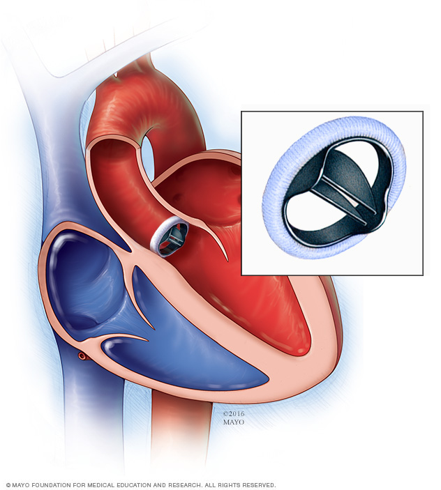
In a mechanical valve replacement, an artificial heart valve made of strong material replaces the damaged valve.
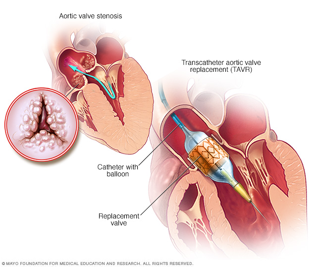
Transcatheter aortic valve replacement (TAVR) is a type of heart valve surgery. It's done to replace a narrowed aortic valve, a condition called aortic valve stenosis. A doctor inserts a flexible tube called a catheter into a blood vessel and guides it into the heart. A replacement valve made of cow or pig tissue goes through the tube to the specific area in the heart. A balloon on the catheter tip inflates to press the new valve into place. Some valves are self-expanding.
Lifestyle and home remedies
If you have aortic valve stenosis, you'll likely need regular health checkups. Continue taking all your medications as prescribed.
Making certain lifestyle changes can help keep the heart healthy and may prevent or slow heart disease. Try these heart-healthy tips:
- Don't smoke. If you smoke, quit. Ask your provider about resources to help you quit smoking. Joining a support group may be helpful.
- Eat a heart-healthy diet. Eat a variety of fruits and vegetables, low-fat or fat-free dairy products, poultry, fish, and whole grains. Avoid saturated and trans fat, and excess salt and sugar.
- Maintain a healthy weight. Aim to keep a healthy weight. Lose weight if you're overweight or have obesity. Losing just a few pounds can help reduce risk factors for heart disease. Talk to your provider about the weight that's best for you.
- Get regular exercise. Exercise helps manage weight and control risk factors for heart disease. Aim to get about 30 minutes of physical activity, such as brisk walks, each day.
- Manage stress. Find ways to help reduce emotional stress. Getting more exercise, practicing mindfulness, spending time with family and friends, and connecting with others in support groups are some ways to reduce stress.
Pregnancy and aortic valve disease
If you have aortic stenosis and are considering pregnancy, it's important to talk to your health care provider about your plans. Together, you and your health care provider can discuss the safety of medications and whether you need a procedure to treat aortic valve stenosis before getting pregnant.
Those with heart valve disease such as aortic valve stenosis usually require close monitoring by a health care provider during pregnancy. Care providers may recommend avoiding pregnancy due to the risk of complications if you have severe aortic stenosis.
Preparing for an appointment
If you think you have aortic valve stenosis, consider being evaluated and treated at a medical center with a multidisciplinary heart valve team. This is a team of heart doctors (cardiologists) and other care providers trained and experienced in evaluating and treating heart valve disease.
Here's some information to help you prepare for your appointment.
What you can do
- Be aware of pre-appointment restrictions. When you make the appointment, ask if there's anything you need to do beforehand.
- Write down your symptoms, including any that seem unrelated to heart valve disease.
- Write down key personal information, including a family history of heart disease and any major stresses or recent life changes.
- Make a list of all medications, vitamins and supplements you take.
- Take a family member or friend along, if possible. Someone who goes with you can help you remember information you receive.
- Be prepared to discuss your diet and exercise habits. If you don't already eat well and exercise, be ready to talk to your health care provider about challenges you might face in getting started.
- Write down questions to ask your care provider.
For aortic valve stenosis, some basic questions to ask your health care provider include:
- What is likely causing my symptoms or condition?
- What are other possible causes for my symptoms or condition?
- What tests will I need?
- What's the best treatment?
- What are the alternatives to the primary approach you're suggesting?
- I have other health conditions. How can I best manage them together?
- Are there restrictions I need to follow?
- Should I see a specialist?
- If I need surgery, which surgeon do you recommend for heart valve surgery?
- Is there a generic alternative to the medicine you're prescribing?
- Are there brochures or other printed material I can take with me? What websites do you recommend?
Don't hesitate to ask other questions you have.
What to expect from your doctor
Your health care provider is likely to ask you a number of questions, including:
- When did your symptoms begin?
- Do you always have symptoms or do they come and go?
- How severe are your symptoms?
- What, if anything, improves your symptoms?
- What, if anything, worsens your symptoms?
Last Updated Aug 18, 2022
© 2024 Mayo Foundation for Medical Education and Research (MFMER). All rights reserved. Terms of Use


